NCLEX EKG Rhythms: A Strip-Reading Workshop
Work through original sinus rhythm, atrial fibrillation and ventricular tachycardia strips while keeping pulse and perfusion central.

A monitor can show the same wide-complex tachycardia in two patients while the immediate response differs: one has a pulse and the other does not. Naming the rhythm is useful, but it does not finish an NCLEX EKG question.
This workshop uses a small set of original synthetic ECG strips to practice observation before action. EKG and ECG refer to the same test. The drawings show selected rhythm features; they are not recordings from patients, validated diagnostic devices or official NCLEX material.
Calibrate the strip before counting
The teaching strips use the conventional scale of 25 mm/second and 10 mm/mV. At that speed, a small horizontal square represents 0.04 second and a large square 0.20 second. Each strip spans six seconds. Screen resizing changes physical dimensions, so use the labeled time grid rather than a ruler held against the display.
For a regular rhythm, 300 divided by the number of large squares between R waves estimates rate. For an irregular rhythm, counting QRS complexes over a known interval and converting to a minute is an estimate; a longer sample is more representative.
Use five observations in order: rate, regularity, P waves, PR relationship and QRS width. The Open RN basic ECG chapter explains these measurements. An adult PR interval is normally 0.12–0.20 second. A QRS of 0.12 second or more is wide; identify the scale before judging it.
Strip A: make the normal pattern explicit
The R waves in the first strip are 0.80 second apart: four large squares. Dividing 300 by four gives 75 beats/minute. The spacing is regular; a similar P wave precedes each QRS, each P is followed by a QRS, and the modeled PR interval is 0.16 second. The QRS lasts 0.08 second.
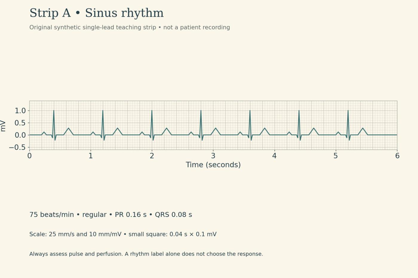
Together, those observations support normal sinus rhythm. “It looks organized” is less useful because several abnormal rhythms also look organized. Record the relationship between atrial and ventricular activity.
Keep the sinus sequence but slow the rate below 60: the pattern becomes sinus bradycardia. Speed it above 100: sinus tachycardia. Those labels do not tell you whether the person is unstable or why the rate changed. A sleeping, comfortable patient and a patient with hypotension and altered mental status need different assessments even if their rates match.
For sinus tachycardia, look for a driver such as pain, fever or volume loss rather than assuming the monitor identifies a primary arrhythmia that needs a rate-slowing intervention. A rate can be a response to illness.
Strip B: the irregular spacing is only the first clue
The second strip has irregular R–R intervals and no consistent discrete P waves. The QRS complexes in this example remain narrow. Those combined findings illustrate atrial fibrillation.
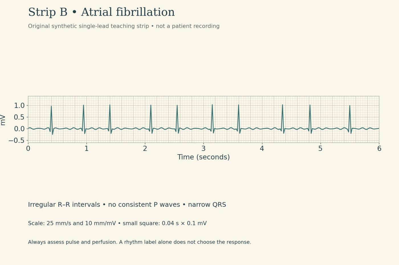
Do not measure a single invented PR interval where there is no consistent P wave to use. Also avoid diagnosing atrial fibrillation from irregularity alone: ectopic beats and artifact can create irregular spacing. Confirm the signal, compare the clinical pulse and obtain the appropriate diagnostic assessment.
Atrial flutter differs: organized atrial activity can produce repeated flutter waves, often described as sawtooth. Ventricular conduction may be regular or variable. Neither atrial fibrillation nor flutter has to produce the same ventricular rate in every patient.
The OpenStax dysrhythmia chapter provides additional rhythm context. A 12-lead ECG and the clinical record can answer questions a short single-lead teaching strip cannot.
A fast narrow rhythm does not identify one mechanism
A regular narrow-complex tachycardia with P waves difficult to identify can suggest a supraventricular tachycardia. “SVT” is a broad category; in many teaching questions it describes a regular rapid narrow-complex presentation. It should not make you ignore a visible sinus P-wave relationship or assume every rapid rhythm is the same disorder.
At the bedside, assess symptoms and perfusion. The 2025 AHA adult advanced life support guidance emphasizes clinical stability and whether the arrhythmia is causing the deterioration. Hypotension, acutely altered mental status, shock, ischemic discomfort or acute heart failure change the urgency.
Strip C: the monitor cannot supply the pulse
The third strip illustrates a regular wide-complex tachycardia at 180 beats/minute, with modeled QRS complexes lasting 0.16 second and no consistent preceding P waves. In the original exercise, the rhythm is monomorphic ventricular tachycardia.
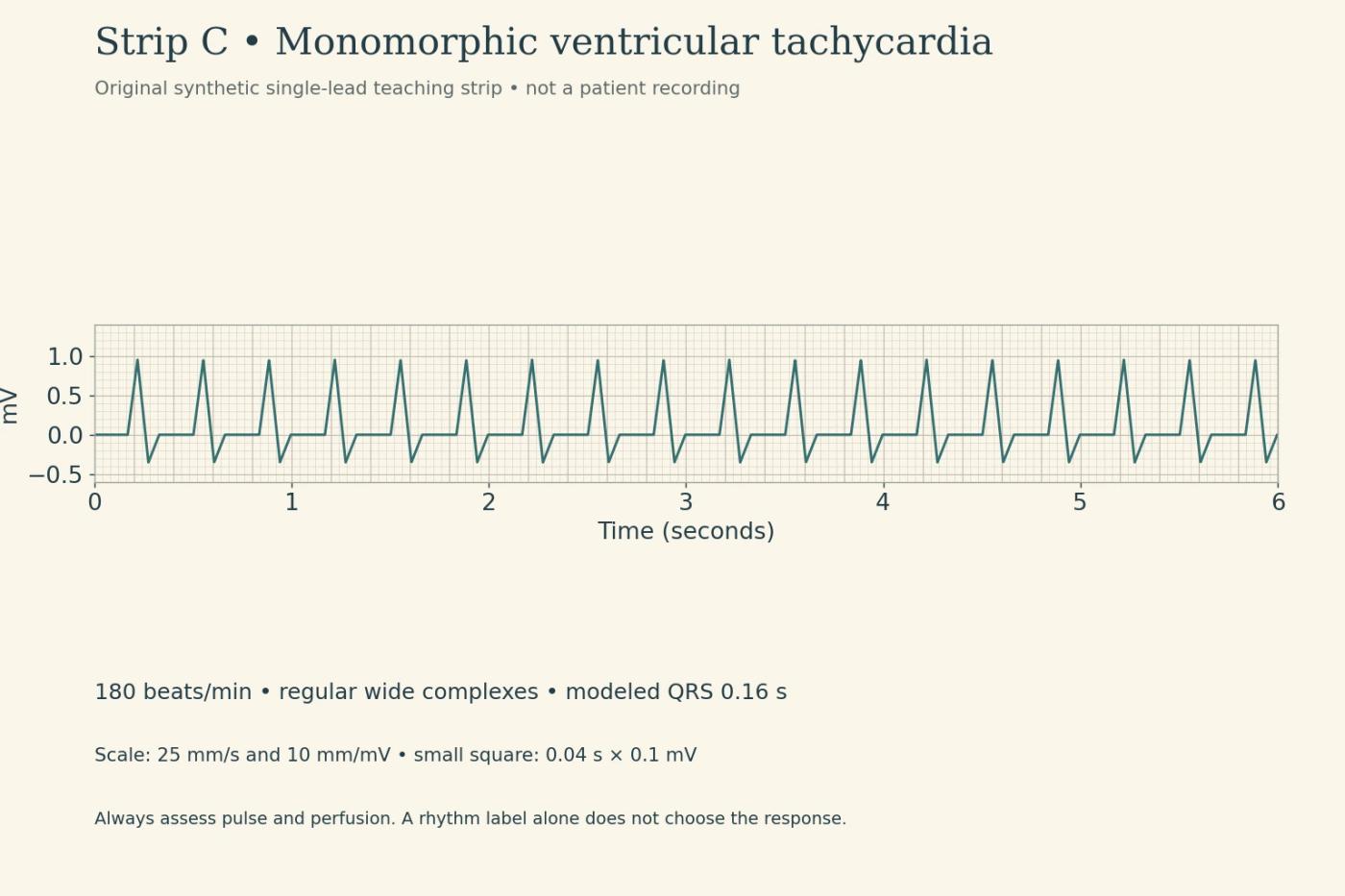
Patient 1 is unresponsive, is not breathing normally and has no definite pulse on the rapid assessment. Activate the emergency response and begin the cardiac-arrest pathway, including high-quality CPR and prompt defibrillation for pulseless VT according to training and protocol.
Patient 2 is awake and has a pulse, but is hypotensive with new confusion. This is a pulse-present unstable tachyarrhythmia situation requiring immediate help and preparation for the appropriate emergency treatment. Do not start chest compressions merely because the strip says VT. Management depends on the rhythm and clinical state; unstable monomorphic VT with a pulse generally requires synchronized cardioversion by the trained team.
The strip is identical. The pulse and perfusion findings change the pathway. This comparison concerns monomorphic VT; it should not be generalized to polymorphic VT, for which the AHA recommends immediate defibrillation.
Chaotic activity and a flat line require different checks
Ventricular fibrillation has disorganized electrical activity without identifiable, organized QRS complexes. In a pulseless patient it is a shockable arrest rhythm. Asystole is absence of discernible ventricular electrical activity; it is not shockable. An organized electrical rhythm without a pulse is pulseless electrical activity, also not shockable.
A flat monitor display still requires checking the patient and the equipment connection and confirming the rhythm as appropriate, without delaying needed resuscitation. Disconnected leads and artifact can mislead. The point is to avoid treating a screen failure as a diagnosis while also avoiding prolonged rhythm analysis in cardiac arrest.
The separate AHA adult basic life support guidance explains rapid recognition and assessment. These paragraphs teach the distinction between pathways; they are not a replacement for certified resuscitation training or a complete medication-and-energy algorithm.
Finish with a bedside report, not just a rhythm name
For each strip, complete this brief report: “The rhythm features are… The patient has or does not have a pulse… The perfusion findings are… The immediate nursing responsibility is…”
If the question supplies an electrolyte result, connect it to the rhythm and the clinical situation rather than treating every abnormal number as the same priority. The laboratory interpretation guide develops that skill. For competing patient needs, use the prioritization comparisons.
In ENDPEAK's RN Topics and QBank, use rhythm review to practice this full report. The next useful task is to hide a strip's label and defend the observation—not to memorize the silhouette alone.
References checked October 2026: Open RN Nursing Advanced Skills ECG chapter; OpenStax Medical-Surgical Nursing; 2025 AHA BLS and ALS guidance. Traces and scenarios are original educational constructions. No independent cardiology or nursing review is claimed.
ENDPEAK publishes independent NCLEX-RN study resources. Exam rules are checked against official NCSBN sources. Original teaching examples are identified as such, and clinical review is credited only when it has actually occurred. ENDPEAK is not affiliated with or endorsed by NCSBN.