NCLEX Fetal Heart Rate Decelerations: Read Timing and Variability
Align fetal heart rate changes with contractions, then use variability and the clinical record to interpret what the pattern means.

Two fetal heart rate dips reach the same depth. One reaches its lowest point with the contraction peak; the other bottoms out afterward. Calling both “a low heart rate” loses the timing that distinguishes early from late decelerations.
Start this visual NCLEX guide by aligning the upper fetal heart rate trace with the lower contraction trace. Then examine the descent, the recovery and the baseline between events. A shape is a clue; the complete maternal–fetal picture determines its significance.
The diagrams are original, mathematically constructed teaching traces—not patient recordings or official examination items. Their time axes are explicit. Short examples illustrate morphology; a clinical baseline and recurrence assessment require the appropriate longer observation window.
Read the space between the dips
Before labeling a deceleration, identify the baseline and variability. Baseline is assessed over a 10-minute window, excluding relevant episodic changes; 110–160 beats/minute is the usual normal range. Variability describes baseline fluctuations: absent means undetectable, minimal is detectable at 5 beats/minute or less, moderate is 6–25 and marked is greater than 25.
Do not count a large deceleration as baseline variability. Likewise, a number inside 110–160 does not make the whole trace normal. A baseline of 140 with recurrent late decelerations and absent variability is a very different situation from 140 with moderate variability and no concerning changes.
Accelerations provide another part of the picture. At 32 weeks or later, the usual definition includes an abrupt rise of at least 15 beats/minute lasting at least 15 seconds but less than two minutes; before 32 weeks, the size and duration criteria are 10 beats/minute and 10 seconds. Absence of accelerations alone does not establish fetal acidemia. Review the basic monitoring terminology alongside the patterns.
Early: put a vertical line through the contraction peak
In the first teaching trace, a contraction begins at 20 seconds, peaks at 60 and ends at 100. The fetal rate decreases gradually over the same interval, with its nadir at 60. The onset-to-nadir interval is 40 seconds.
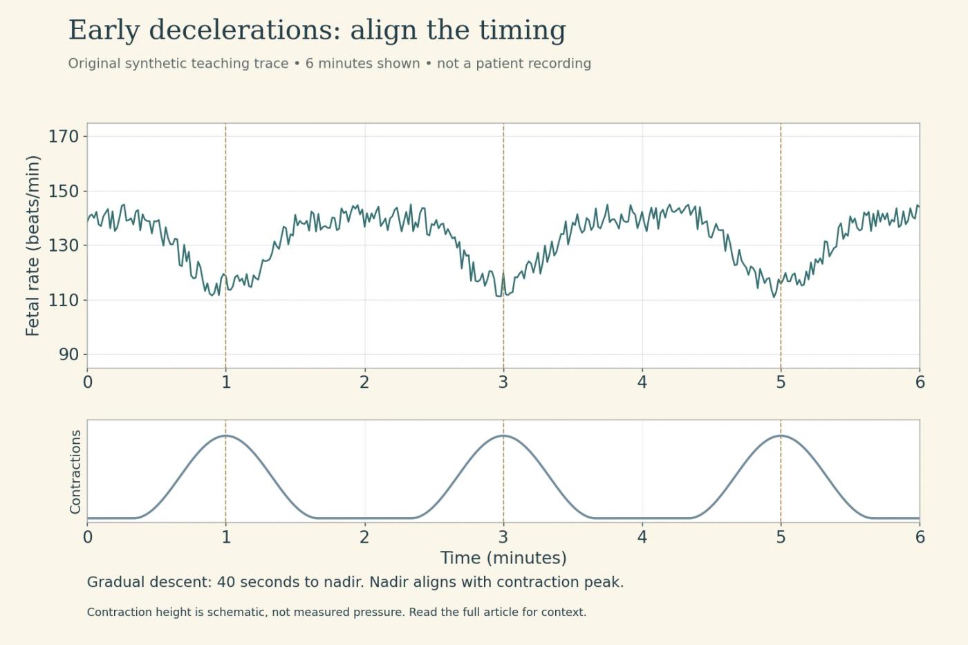
That timing illustrates an early deceleration: a gradual decrease and recovery that roughly mirror the contraction. Fetal head compression is the classic association. The useful visual relationship is the shared timing, not merely a rounded shape.
With otherwise reassuring features, an early deceleration generally does not call for a corrective maneuver directed at the dip itself. Continue appropriate assessment and routine care. If other features become concerning, do not let the early label overrule them.
Late: the nadir moves to the right
Keep the contraction in the same position. In the second trace, the gradual dip begins at 60 seconds, reaches its nadir at 100 and returns to baseline at 140. The nadir follows the contraction peak, and recovery follows the contraction's end.
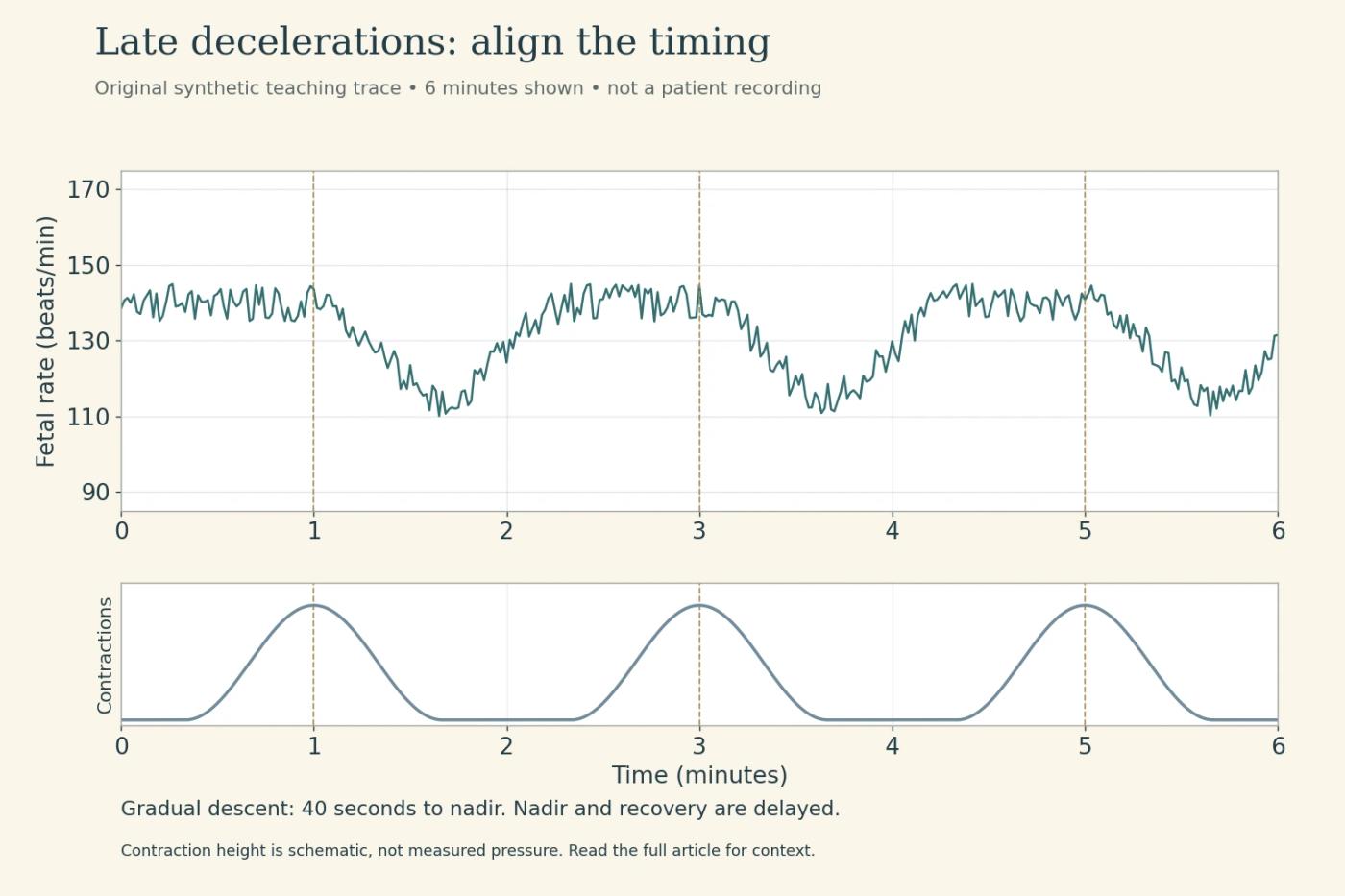
This is the delayed relationship characteristic of a late deceleration. It raises concern about oxygen transfer through the uteroplacental circulation. It does not, by itself, identify the exact maternal or placental cause or prove a particular degree of fetal acidemia.
The comparison is easier when you mark three events on both traces: onset, peak or nadir, and recovery. A student who looks only for a U shape can miss the delay. The MSD professional monitoring reference describes these temporal relationships.
Variable: inspect the speed of the descent
The third teaching trace has dips with changing timing relative to successive contractions. Each falls abruptly; onset to nadir is less than 30 seconds. The standard variable-deceleration definition also requires a decrease of at least 15 beats/minute lasting at least 15 seconds and less than two minutes.
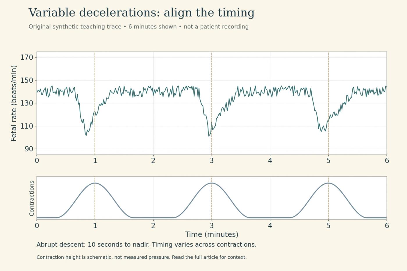
Cord compression is the usual association. A V, U or W outline can appear, but a letter shape is not the definition. Look at abruptness, depth, duration, recovery and how often the events occur.
Do not treat every variable deceleration as equivalent. An isolated event with prompt recovery and moderate variability is not the same picture as recurrent events with slow recovery and loss of variability. A sustained decrease lasting two minutes or more but less than 10 minutes belongs to the prolonged-deceleration definition; a change persisting 10 minutes is a baseline change.
Exercise: the late dips match, but the assessment does not
Compare the paired diagrams. Both show a baseline near 140 and the same delayed dips. In the fictional clinical record accompanying each illustration, late decelerations occur with more than half of contractions over the preceding 20 minutes.
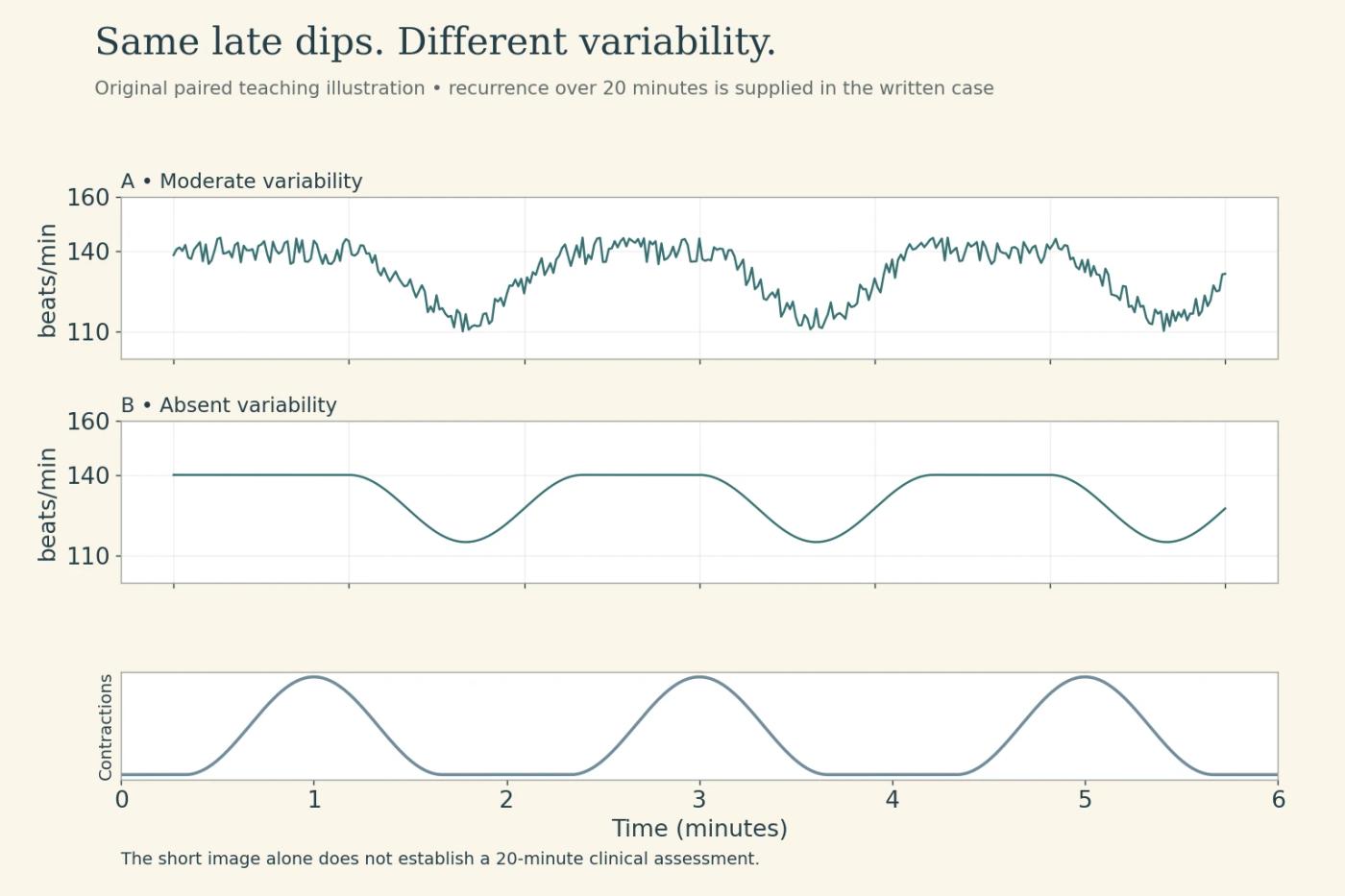
Record A: Moderate variability remains between events. The mother is alert; a recent blood-pressure reduction after analgesia is being assessed.
Record B: Baseline variability is absent, and the recurrent late decelerations continue. Initial corrective measures have not restored the pattern.
Record A fits category II, requiring evaluation and appropriate measures with reassessment. Record B has the combination of absent variability and recurrent late decelerations that meets category III criteria and requires urgent team evaluation and response. Under ACOG's 2025 guidance, a category III pattern that does not respond to initial intrauterine resuscitative attempts calls for expedited delivery when indicated.
The exercise changes one crucial feature while holding the dip timing constant. Neither strip should be interpreted by its lowest number alone. Short drawings cannot establish the full observation history; that history is explicitly supplied in the scenario.
Connect a concerning pattern to a cause you can assess
Check the signal and compare the maternal pulse if there is doubt about which heart rate is being recorded. Assess maternal vital signs, oxygenation, uterine activity, medications and recent events. An external contraction sensor provides timing information; its displayed height should not be treated as a calibrated measure of contraction strength.
For category II patterns, current ACOG guidance supports measures selected for the situation, such as maternal repositioning, addressing hypotension, adjusting induction or augmentation agents, ordered fluid therapy or amnioinfusion when appropriate. These are not interchangeable instructions for every deceleration. Follow the clinical assessment, orders, scope and local protocol, and evaluate the response.
Routine maternal oxygen solely for category II or III tracings is not recommended when maternal hypoxia is absent. That is a particularly important correction to older memory lists. Maternal hypoxia still requires appropriate assessment and treatment.
The current reference is ACOG Clinical Practice Guideline No. 10, October 2025. It replaces older monitoring bulletins and the 2022 oxygen advisory. A university-hosted copy of the guideline provides the full source. The OpenStax nursing discussion also explains checking the maternal versus fetal signal.
What VEAL CHOP leaves out
The mnemonic connects variable with cord compression, early with head compression, accelerations with a generally reassuring response and late with a uteroplacental concern. It does not tell you the baseline, variability, recurrence, maternal condition or response to an intervention.
Use it after recognizing the pattern, not as a substitute for reading the record. The NGN case-study guide can help organize what changes as new findings arrive; the RN test plan overview places maternal–newborn decisions within broader preparation.
Before leaving a practice trace, give one sentence of evidence: “The nadir follows the contraction peak, and variability between recurrent events is…” If the sentence stops before variability, the interpretation is unfinished.
Sources checked October 2026. Original teaching traces and scenarios; no independent obstetric professional review is claimed. This educational interpretation guide does not replace fetal-monitoring training or a patient-specific management plan.
ENDPEAK publishes independent NCLEX-RN study resources. Exam rules are checked against official NCSBN sources. Original teaching examples are identified as such, and clinical review is credited only when it has actually occurred. ENDPEAK is not affiliated with or endorsed by NCSBN.